
+918929672099
Call UsWhat is Bronchogenic Carcinoma? Causes, Types and Diagnosis

Dr. Vrundali Kannoth|5 min read|
When doctors use the term bronchogenic carcinoma, they're referring to the most common form of lung cancer. This medical terminology can feel overwhelming when you're trying to understand a diagnosis.
Breaking down complex medical terms helps. Understanding what is bronchogenic carcinoma helps you to ask informed questions and participate actively in treatment decisions.
This comprehensive guide explains the disease's origins, causes, types, symptoms, and diagnostic process.
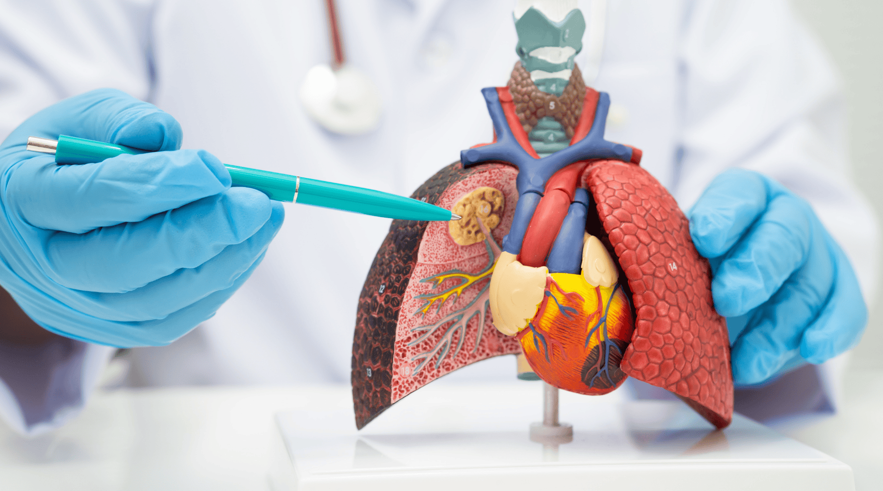
What is bronchogenic carcinoma?
Bronchogenic carcinoma meaning refers to malignant tumours originating in the bronchial epithelial cells lining the airways. The term breaks down as "broncho" (airways), "genic" (arising from), and "carcinoma" (cancer of epithelial tissue).
According to research , bronchogenic carcinoma lung cancer represents approximately 90% of all lung cancers. The remaining 10% includes mesothelioma and other rare lung tumours.
Origins in bronchial epithelial cells
Your airways are lined with epithelial cells that produce mucus and help clear debris. When these cells' DNA becomes damaged by carcinogens, substances that cause cancer, they may transform into malignant cells.
These abnormal cells divide uncontrollably, forming tumours that invade surrounding tissues. Unlike benign growths, cancer cells can spread to lymph nodes and distant organs through the bloodstream or lymphatic system.
Primary bronchogenic carcinoma develops through accumulated genetic mutations over time. Typically, multiple DNA changes must occur before normal cells become cancerous.
Causes and risk factors
Understanding bronchogenic carcinoma causes helps identify who's at highest risk and guides prevention strategies.
1. Smoking
Tobacco smoking remains the leading cause of bronchogenic carcinoma. According to the World Health Organization, smoking causes 85-90% of lung cancer cases globall
Cigarette smoke contains over 70 known carcinogens. These chemicals directly damage bronchial cell DNA, triggering mutations that can lead to cancer. Risk increases with pack-years (cigarettes per day × years smoking).
2. Secondhand smoke exposure
Non-smokers exposed regularly to tobacco smoke face an increased risk. The International Agency for Research on Cancer classifies secondhand smoke as a Group 1 carcinogen, a definite cancer-causing agent.
3. Asbestos exposure
Occupational asbestos exposure significantly raises bronchogenic carcinoma risk factors. Workers in construction, shipbuilding, and manufacturing face elevated risk. Combined smoking and asbestos exposure multiplies risk exponentially.
4. Air pollution
Outdoor air pollution, particularly fine particulate matter (PM2.5), contributes to lung cancer risk. Urban residents in highly polluted areas show higher incidence rates.
Indoor pollution from cooking fuels also contributes significantly in developing regions.
5. Radon gas
This naturally occurring radioactive gas seeps from underground into homes. Radon exposure is the second leading cause of lung cancer after smoking. Testing homes for radon levels is recommended.
6. Genetic factors
Family history of lung cancer increases risk, suggesting inherited susceptibility. Certain genetic variations affect how efficiently the body repairs DNA damage or metabolises carcinogens.
7. Previous lung disease
Chronic obstructive pulmonary disease, Interstitial Lung Disease, and tuberculosis scarring all elevate risk. Chronic inflammation and tissue damage create environments favouring cancer development.
Types and classification of bronchogenic carcinoma
Classification of bronchogenic carcinoma divides tumours into two major categories based on microscopic appearance. This classification drives treatment decisions.
Non-small cell lung carcinoma (NSCLC)
Non small cell lung cancer comprises approximately 85% of bronchogenic carcinoma types. It grows more slowly than small cell type, allowing more treatment options when detected early.
NSCLC subdivides into three main types:
1. Adenocarcinoma:
Adenocarcinoma lung is the most common subtype, representing 40% of lung cancers. It originates in mucus-producing cells lining the airways' outer regions.
This type occurs more frequently in non-smokers and women compared to other subtypes. It typically grows in the lung periphery rather than the central airways.
2. Squamous cell carcinoma:
Squamous cell lung carcinoma accounts for 25-30% of lung cancers. It develops in flat cells lining the airways' inner surfaces.
This type strongly associates with smoking history. Tumours typically arise centrally near major bronchi. They often cause airway obstruction and respiratory symptoms earlier than peripheral adenocarcinomas.
3. Large cell carcinoma:
This aggressive subtype represents 10-15% of NSCLCs. Cells appear large and abnormal under microscopy, but don't fit adenocarcinoma or squamous cell patterns.
Large cell carcinomas can occur anywhere in the lungs. They tend to grow and spread quickly, often presenting at advanced stages.
Small cell lung carcinoma (SCLC)
Small cell lung cancer comprises approximately 15% of cases. It's one of the most aggressive lung cancer types, growing and spreading rapidly.
SCLC almost exclusively affects smokers. Tumours typically arise centrally near major airways. Cancer often spreads to lymph nodes and distant organs before diagnosis.
This type responds well to chemotherapy initially but frequently recurs. Five-year survival remains low despite treatment advances.
Signs and symptoms
Bronchogenic carcinoma signs and symptoms often appear late, contributing to delayed diagnosis. Early-stage disease frequently causes no symptoms.

Early symptoms may include:
- •Persistent cough lasting more than 3 weeks
- •Changes in chronic cough pattern
- •Coughing up blood (haemoptysis), even small amounts
- •Shortness of breath with usual activities
- •Chest pain that worsens with breathing, coughing, or laughing
- •Hoarseness or voice changes
- •Wheezing
- •Recurrent respiratory infections (pneumonia, bronchitis)
Advanced bronchogenic carcinoma symptoms include:
- •Significant unexplained weight loss (>5 kg)
- •Severe fatigue and weakness
- •Loss of appetite
- •Bone pain if metastasised to skeleton
- •Headaches if brain metastases present
- •Yellowing skin and eyes if liver involved
- •Swollen lymph nodes in the neck or above the collarbone
Paraneoplastic syndromes:
Some bronchogenic carcinomas produce hormones or immune responses, causing symptoms unrelated to tumour location. These include elevated calcium levels, low sodium, or neurological symptoms.
Lung cancer symptoms vary significantly between individuals. Persistent respiratory symptoms warrant medical evaluation, especially in high-risk individuals.
Diagnosis of bronchogenic carcinoma
Bronchogenic carcinoma diagnosis involves multiple steps to confirm cancer presence, determine type, and assess spread.
Imaging studies
- •Chest X-rayOften the first test performed. It can identify suspicious masses or nodules but lacks detail for definitive diagnosis.
- •CT scanProvides detailed cross-sectional images showing tumour size, location, and lymph node involvement. Contrast-enhanced CT reveals tumour characteristics and blood vessel relationships.
- •PET scanUses radioactive glucose to identify metabolically active cancer cells. It's excellent for detecting bronchogenic carcinoma metastasis to lymph nodes or distant organs.
- •MRIParticularly useful for brain imaging when metastases are suspected or for detailed evaluation of tumour invading the chest wall.
Tissue diagnosis
Imaging suggests cancer, but tissue confirmation is essential for lung cancer diagnosis.
- •BronchoscopyA thin, flexible tube with a camera passes through the airways. Doctors visualise tumours and obtain bronchogenic carcinoma specimens through biopsy forceps or brushings.
- •CT-guided needle biopsyFor peripheral tumours inaccessible by bronchoscopy, radiologists use CT guidance to insert needles through the chest wall, obtaining tissue samples.
- •Sputum cytologyExamining coughed-up mucus under microscopy can sometimes reveal cancer cells. This non-invasive method works best for central tumours shedding cells into the airways.
- •Surgical biopsyWhen other methods fail, video-assisted thoracoscopic surgery or open biopsy provides definitive tissue diagnosis.
Molecular testing
Modern lung cancer diagnosis includes genetic testing of tumour tissue. Identifying specific mutations (EGFR, ALK, ROS1, KRAS) guides targeted therapy selection. PD-L1 testing predicts immunotherapy response.
Staging
Bronchogenic carcinoma staging uses the TNM system: Tumour size and invasion (T), lymph Node involvement (N), and Metastasis presence (M). Stages range from I (localised, small tumour) to IV (distant spread).
Accurate staging requires integrating imaging, biopsy, and sometimes surgical exploration findings.
Complications and prognosis
Accurate staging requires integrating imaging, biopsy, and sometimes surgical exploration findings.
Physical complications include:
- •Airway obstruction causing breathlessness or collapse
- •Respiratory failure requiring mechanical ventilation
- •Superior vena cava syndrome (facial swelling, breathing difficulty)
- •Pleural effusion (fluid around lungs)
- •Haemoptysis potentially causing severe bleeding
- •Recurrent infections from blocked airways
Bronchogenic carcinoma prognosis depends on multiple factors:
- •Cancer stage at diagnosis (most critical factor)
- •Tumour type (NSCLC generally better than SCLC)
- •Molecular characteristics affecting treatment options
- •Patient's overall health and ability to tolerate treatment
- •Response to initial therapy
Is bronchogenic carcinoma curable?
Early-stage disease offers cure potential through surgery combined with adjuvant therapy. Advanced disease focuses on prolonging life and maintaining quality rather than cure.
Treatment options
Bronchogenic carcinoma treatment approaches depend on cancer type, stage, molecular characteristics, and patient factors. Your oncology team tailors therapy to your specific situation, often combining multiple approaches for optimal outcomes.
- •Surgery:Cancer treatment for early-stage NSCLC preferably involves surgical removal. Minimally invasive video-assisted thoracoscopic surgery offers faster recovery than traditional open approaches.
- •Radiation therapy:External beam radiation targets tumours with high-energy rays, used for patients unable to tolerate surgery or as adjuvant treatment after surgery.
- •Chemotherapy:Systemic chemotherapy uses drugs killing rapidly dividing cells, with platinum-based regimens standard for both NSCLC and SCLC.
- •Targeted therapy:For tumours with specific mutations, targeted drugs block pathways driving cancer growth.
- •Immunotherapy:Checkpoint inhibitors help immune systems recognise and attack cancer cells. They've revolutionised treatment for advanced NSCLC, significantly extending survival in responding patients.
Conclusion
Bronchogenic carcinoma represents a serious diagnosis requiring prompt, comprehensive evaluation. Understanding the disease helps you participate actively in treatment decisions.
Early detection dramatically improves outcomes. If you're at high risk, a current or former smoker, occupational exposures, or have a family history, discuss screening with your doctor. Low-dose CT screening can detect cancers when they are still curable through surgery.
Treatment advances continue to improve survival rates, particularly for patients with targetable mutations or those responding to immunotherapy. Even advanced disease now has more options than ever before.
Don't hesitate to seek second opinions or ask questions about your care. Connect with specialist oncology teams experienced in managing bronchogenic carcinoma for comprehensive evaluation and personalised treatment planning.
FAQs
Bronchogenic carcinoma is essentially synonymous with lung cancer, representing approximately 90% of all lung malignancies. When doctors say "lung cancer," they're usually referring to bronchogenic carcinoma.
Related Blogs

Symptoms6 min read
Skin Clues You Shouldn’t Ignore: Rare Lung Cancer Symptoms on Skin
Dr. Vrundali Kannoth
Prevention5 min read
Air Pollution and Lung Cancer: Risks, Progression, and What Patients Should Do
Dr. Vrundali Kannoth