
+918929672099
Call UsFIGO Staging Ovarian Cancer: Guide to Stages & Their Meaning
Dr. Vrundali Kannoth|5 min read|
When someone is told they have ovarian cancer, it’s not just a diagnosis; it’s the beginning of a hundred emotions and questions.
The medical terms that follow sound distant, but one of the most important among them is FIGO staging of ovarian cancer.
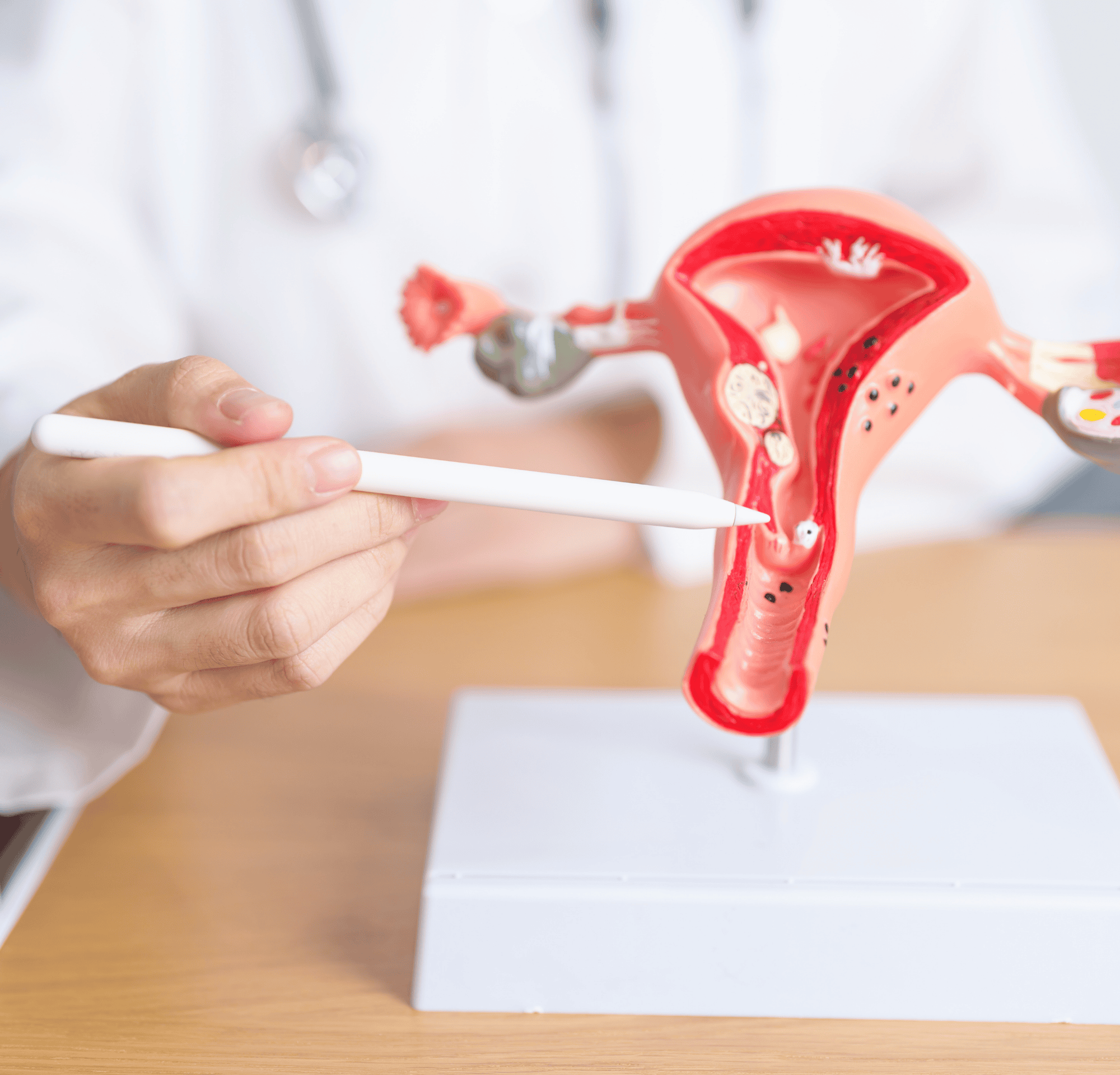
The ovaries have a big role in producing hormones and supporting fertility. When cancer begins there, doctors rely on the latest FIGO staging of ovarian cancer to understand how far it has spread.
Let’s take a closer look at what the FIGO staging system for ovarian cancer actually means and how it connects to treatment and recovery.
What is FIGO staging of ovarian cancer?
The FIGO staging of ovarian cancer system, created by the International Federation of Gynaecology and Obstetrics (FIGO), classifies ovarian cancer based on how far it has spread.
Essentially, it shows whether the cancer is contained within the ovaries or has moved to other parts of the body.
The FIGO classification of ovarian cancer relies on information from surgery, imaging, and pathology. By combining these details, doctors can assign a precise stage, from Stage I (limited to the ovary) to Stage IV (spread to distant organs).
Following the FIGO guidelines of ovarian cancer ensures that healthcare professionals around the world approach diagnosis and treatment consistently.
Breakdown of FIGO staging of ovarian cancer
Understanding the FIGO staging system for ovarian cancer helps put medical details into perspective.
Stage I
- •tage IA:In FIGO stage 1A, ovarian cancer is found in a single ovary (or fallopian tube) and has not spread.
- •Stage IB:Both ovaries (or tubes) are affected but contained within them.
- •Stage IC:Cancer remains limited to the ovaries but shows higher risk features such as: 1. rupture of the outer capsule before or during surgery, or 2. cancer cells detected in abdominal fluid (ascitic fluid or washings)
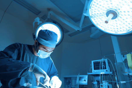
Treatment:
Surgery is curative at this stage. Doctors remove the ovaries, fallopian tubes, and uterus, followed by examination of nearby tissues to ensure no microscopic spread. Chemotherapy may be advised in Stage IC for added safety.
Outlook:
Stage I ovarian cancer has one of the best survival rates, and early detection can lead to full recovery with routine follow-ups.
Stage II
- •Stage IIA:The tumour has spread from the ovaries to nearby reproductive organs such as the uterus or fallopian tubes.
- •Stage IIBThe spread involves other pelvic structures like the bladder, rectum, or pelvic wall.
Treatment
A combination of surgery and chemotherapy is recommended. The surgical aim is to remove all visible cancer, followed by chemotherapy to destroy any remaining microscopic cells.
Outlook:
With timely intervention, many patients respond well.
Stage III
- •Stage IIIA:Microscopic cancer cells are detected outside the pelvis, usually on the abdominal lining or in nearby lymph nodes.
- •Stage IIIB:Visible tumour deposits (≤2 cm) are found in the abdomen or on the surface of the liver or spleen.
- •Stage IIIC:Larger tumour deposits (>2 cm) or spread to lymph nodes in the abdomen or groin, this is often referred to as FIGO Stage 3C ovarian cancer.
Treatment:
Extensive (cytoreductive) surgery to remove as much tumour as possible, followed by multiple cycles of chemotherapy. In some cases, targeted therapies or maintenance treatments are added to prevent recurrence.
Outlook:
Many patients achieve remission with modern therapy. Care focuses equally on treatment success and maintaining quality of life through care and regular monitoring.
Stage IV
- •Stage IVA:Cancer cells are found in fluid around the lungs (pleural effusion).
- •Stage IVB:Spread to organs outside the abdomen, such as the liver tissue, lungs, or distant lymph nodes.
Treatment:
Treatment is systemic, involving chemotherapy, targeted drugs, and occasionally surgery to manage ovarian cancer symptoms or reduce tumour burden. The focus is on controlling the disease, easing symptoms, and maintaining comfort.
Outlook:
Even at this advanced stage, many women live active lives through treatment and follow-up support. Advances in precision medicine continue to bring better control over the disease’s course.
FIGO staging and ovarian cancer radiology
Imaging plays an essential role in the FIGO staging ovarian cancer radiology process. Radiology helps doctors map the tumour, understand its size and spread, and guide treatment decisions.
Role of CT, MRI, PET scans
Imaging scans play a crucial role in understanding how ovarian cancer has spread. They help doctors plan treatment and monitor disease more accurately.
CT scan (Computed Tomography)
A CT scan gives doctors a broad, three-dimensional view of the abdomen and pelvis. It helps identify the tumour’s size, whether it has spread to nearby organs, and if lymph nodes appear enlarged.
MRI (Magnetic Resonance Imaging)
MRI offers a detailed look at soft tissues in the pelvis. It helps clarify where exactly the tumour is and whether it’s pressing against or invading nearby structures.
This level of precision is especially useful when planning surgery or assessing complex cases where the tumour borders are unclear on a CT scan.
PET scan (Positron Emission Tomography)
PET scans detect metabolic activity: areas where cells are consuming more energy than normal, often a sign of cancer.
By revealing hidden or small metastases that might not be visible on CT or MRI, PET scans provide an extra layer of certainty in staging and treatment planning.
Imaging vs. surgical staging
Imaging offers an initial overview, but only surgical staging can pinpoint the cancer’s precise stage.
| Aspect | Imaging | Surgical staging |
|---|---|---|
| Purpose | Creates a visual roadmap of the tumour and possible spread. | Confirms the exact FIGO stage through direct examination and tissue sampling. |
| Accuracy | Estimates disease extent based on what can be seen on scans. | Provides definitive results by viewing organs directly and testing tissue samples. |
| Use in treatment planning | Helps plan the surgical approach and detect distant metastases before surgery. | Finalises staging, guiding chemotherapy or targeted therapy decisions post-surgery. |
| Limitations | May miss microscopic disease or early spread. | More invasive but offers complete diagnostic clarity. |
FIGO staging and ovarian cancer treatment
FIGO staging ovarian cancer treatment helps doctors decide how extensive surgery should be, and whether chemotherapy or targeted therapy is needed.
How is treatment guided by FIGO stage of ovarian cancer
Ovarian cancer treatment by stage is easier as it makes sure a standard procedure gets followed:
- •Stage I:Surgery is usually enough. In some cases (like Stage IC), short chemotherapy cycles are added for extra safety.
- •Stage II:Surgery followed by chemotherapy to clear both visible and microscopic types of cancer cells.
- •Stage III:Extensive (cytoreductive) surgery combined with several chemotherapy cycles, sometimes followed by maintenance therapy.
- •Stage IV:Systemic treatments such as targeted therapy, immunotherapy, or palliative surgery; aimed at control, comfort, and maintaining quality of life.
Early vs. advanced stages
Treatment approaches differ depending on whether ovarian cancer is detected early or at an advanced stage.
| Stage group | Treatment goal | Treatment plan | What to expect |
|---|---|---|---|
| Early stages (I-II) | Cure the disease completely | Surgery to remove affected organs, sometimes followed by short chemotherapy cycles. | High success rates with regular monitoring after treatment. |
| Advanced stages (III-IV) | Control the disease and extend life | Chemotherapy to shrink tumours, then surgery if possible; may include targeted or maintenance therapy. | Focus on long-term control, symptom relief, and quality of life. |
FIGO staging - ovarian cancer prognosis
FIGO staging of ovarian cancer prognosis helps doctors predict recovery chances and plan treatment intensity with greater precision and compassion.
Survival outlook per stage
- •Stage I:5-year survival often exceeds 90% with timely treatment.
- •Stage II:Around 70%, depending on tumour grade and surgical success
- •Stage III:Typically 40–50%, though outcomes improve with modern therapies and maintenance treatments.
- •Stage IV:About 20–25%, but new targeted options continue to extend survival and quality of life.
Importance of accurate FIGO staging of ovarian cancer
Precise staging ensures that every patient receives care that fits their condition, avoiding both overtreatment and undertreatment. It also helps doctors predict recurrence risk.

According to a 2015 survival analysis validating the accuracy of the revised 2013 FIGO staging system for ovarian cancer, the updated classification more precisely differentiated survival outcomes particularly for patients in Stages I and III.
Mnemonic for FIGO staging of ovarian cancer
Remembering the FIGO staging of ovarian cancer can be simplified with this easy mnemonic:
“O-P-A-L”
- •O -Ovary only (Stage 1)
- •P -Pelvis involved (Stage 2)
- •A -Abdomen spread (Stage 3)
- •L -Lungs/liver distant (Stage 4)
What this means for you
Understanding FIGO staging of ovarian cancer helps you see the disease clearly and makes treatment plans less overwhelming. Each stage shows how your care will be tailored, what treatments may be needed, and what to expect along the way.
Pro tip:
Keep a copy of your staging report and imaging results. Having these details on hand makes it easier to track progress, ask informed questions, and feel more in control of your journey.
With oncology doctors guiding treatment decisions and clarifying FIGO stage of ovarian cancer, patients can approach ovarian cancer with understanding and confidence.
FAQs on FIGO staging of ovarian cancer
Radiology can provide an estimated FIGO staging of ovarian cancer, but the definitive stage is confirmed only through surgical evaluation and pathology.
Related Blogs
Diagnosis5 min read
Ultrasound Kya Hota Hai? (अल्ट्रासाउंड): प्रक्रिया, रिपोर्ट और उपयोग
Dr. Vrundali Kannoth