
+918929672099
Call UsPathophysiology of Ovarian Cancer Explained: Complete Overview
Dr. Vrundali Kannoth|6 min read|
No one walks into a clinic expecting to leave with the label of ovarian cancer. Patients often wonder: Why didn’t I notice anything earlier? Why does this disease progress silently? Or, what could be the best line of treatments for my condition? The answers lie in the pathophysiology of ovarian cancer: what’s happening at the cellular level, why certain symptoms appear, and how the disease moves through the body.
This isn’t just for doctors or scientists.
Learning about ovarian cancer pathophysiology can give you context for your own experience, helping to demystify scans, blood tests, and treatment plans. It can also show why some ovarian cancer types act differently, why complications like ascites develop, and why specific therapies are chosen.
In essence, it turns confusion into clarity, making it easier to have meaningful conversations with your care team.
Theories of Pathogenesis
Before diving deep, it’s useful to know how scientists think ovarian cancer starts. These theories help explain the origins of epithelial ovarian cancer pathophysiology and why some cells turn rogue.
Incessant Ovulation Theory
This theory suggests that repeated ovulatory cycles take a small toll on the ovarian surface. Each time an egg is released, the ovarian epithelium experiences minor injury. Normally, the body repairs this tissue, but over many cycles, DNA mistakes can accumulate, increasing the risk of malignant transformation. This process is a central concept in ovarian tumour pathophysiology, helping explain why factors that reduce ovulation, like pregnancy or long-term oral contraceptive use, can lower ovarian cancer risk.
Fallopian Tube Theory (STIC Lesions)
Areas known as STIC lesions are believed to give rise to high-grade serous tumours. As evidenced by studies identifying the clonal relationship between TIC and metastatic tumours, most serous tumours likely originate in the distal fallopian tube.
These small dysplastic areas eventually become malignant and, due to their strategic location, metastasise to the ovaries and surrounding pelvic structures. Understanding this process is a crucial part of epithelial ovarian cancer pathophysiology and has influenced preventive strategies.
Two-Pathway Model (type I vs type II)
The two-pathway model divides the pathophysiology of ovarian cancer into Type I and Type II, reflecting different biological behaviours. Type I tumours, including low-grade serous and endometrioid cancers, progress slowly and usually arise from well-defined precursor lesions. Type II tumours, like high-grade serous carcinomas, are aggressive, genetically unstable, and often linked to early TP53 mutations.
Molecular Pathophysiology of Ovarian Cancer
Let’s zoom in on what’s happening at the cellular and genetic level: this is where the “why” behind pathophysiology of ovarian cancer comes alive.
Genetic Alterations (TP53, BRCA, ARID1A, KRAS)
Think of your genes as the body’s instruction manual. In ovarian cancer, certain “instructions” get damaged. Check below the mutations and their specifications:
- •TP53Found in high-grade tumours, acts like a broken security system as cells that should die keep multiplying
- •BRCA 1/2Weakens DNA repair
- •ARID1AAppears in clear cell and endometrioid tumours
- •KRASCommon in low-grade tumours, pushes cells into continuous growth mode
Tumour Microenvironment and Immune Escape
Cancer doesn’t develop in isolation. Around the tumour are supporting cells, blood vessels, and immune cells: this is called the ‘tumour microenvironment’. Instead of fighting the cancer, some immune cells get “tricked” into protecting it. The tumour also releases signals that shut down normal immune responses, a process known as ‘immune escape’. That’s why many ovarian tumours can grow undetected for so long, making epithelial ovarian cancer pathophysiology especially complex.
Angiogenesis and Tumour Progression
For a tumour to grow beyond a tiny cluster, it needs food and oxygen. To get these, it creates new blood vessels: a process called ‘angiogenesis’. The key player here is VEGF, which increases blood supply to the tumour. However, VEGF also makes blood vessels leaky, which contributes to fluid buildup in the abdomen, better known as ‘ascites’. This link between angiogenesis and the pathophysiology of ovarian cancer explains why certain drugs, like VEGF inhibitors, are designed to cut off the tumour’s blood supply.
Pathophysiology of Ascites in Ovarian Cancer
If you’ve heard of ascites, fluid buildup in the abdomen, it’s a common problem in ovarian cancer.
The pathophysiology of ascites in ovarian cancer explains why this happens and how it affects symptoms.
VEGF-driven Vascular Permeability
One of the main culprits is VEGF (vascular endothelial growth factor). This molecule makes tiny blood vessels leaky, allowing fluid to escape into the abdominal cavity.
Lymphatic Obstruction
Normally, lymphatic channels drain away excess fluid. But when tumour cells invade or press against these vessels, drainage is blocked. This disruption worsens fluid retention and represents another pathway within ovarian tumour pathophysiology that explains malignant ascites.
Cytokine Involvement
It’s not just about blocked vessels; chemical messengers called cytokines (like IL-6 and IL-8) also play a role. They trigger inflammation and change how surrounding tissues behave, further promoting fluid leakage. This makes the pathophysiology of ascites in ovarian cancer more complex than a single mechanism.
Clinical Impact of Malignant Ascites
Ascites aren't just uncomfortable; they can cause bloating, early satiety, restricted breathing, abdominal pain, and even limit mobility. By grasping the pathophysiology of ascites in ovarian cancer, doctors can better tailor interventions, like paracentesis, diuretics, or targeted drugs, to reduce fluid and ease symptoms.
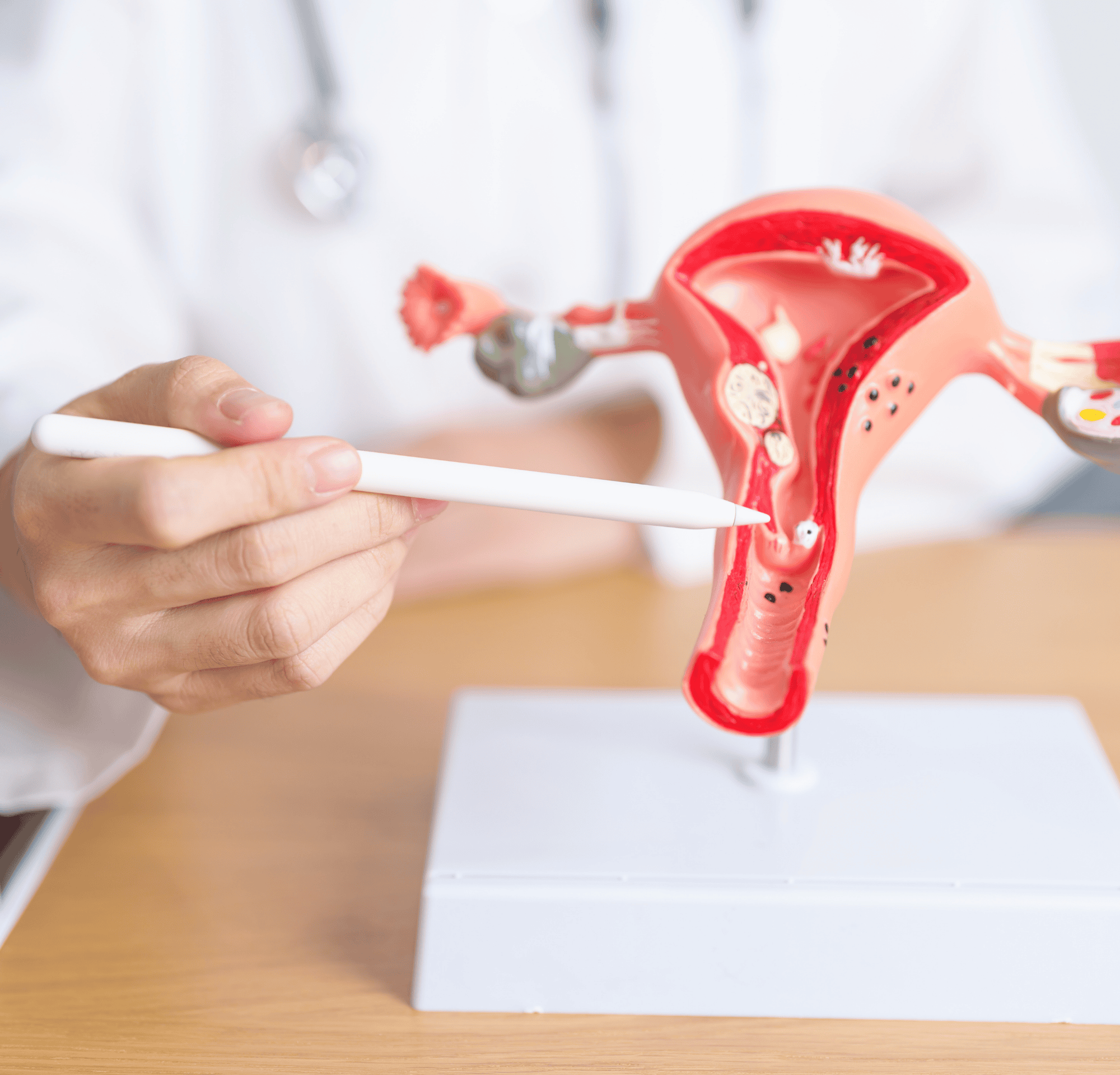
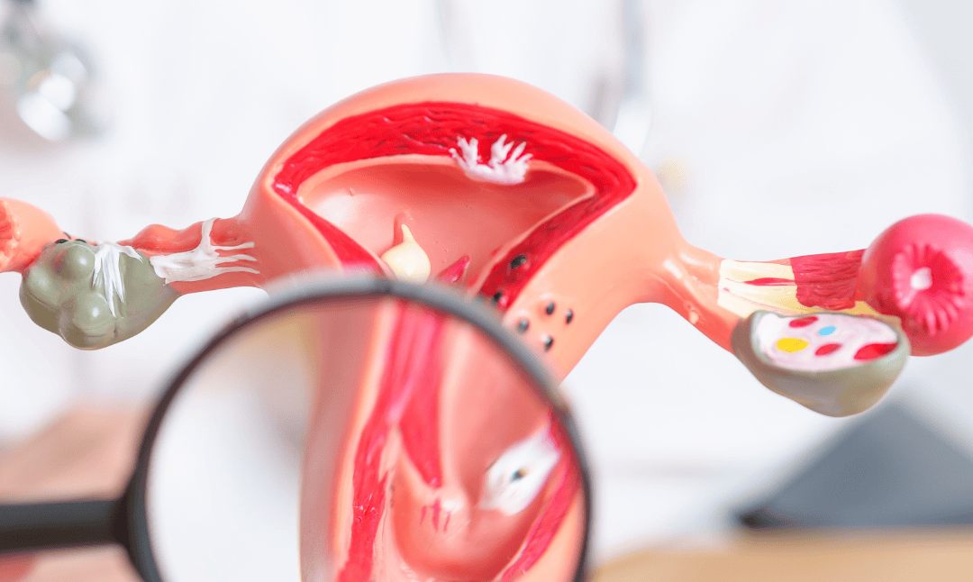
Patterns of Spread and Metastasis
Understanding how ovarian cancer spreads is part of ovarian cancer pathophysiology and guides treatment strategies.
Peritoneal Dissemination
Tumour cells shed from the ovary and implant on the peritoneum (the lining of the abdomen) and the omentum (a fatty apron of tissue). This explains why surgeons aim to remove visible disease during debulking surgery - an important principle of epithelial ovarian cancer pathophysiology.
Lymphatic Routes
Tumour cells also travel through lymph nodes, especially in the pelvis and para-aortic region. This is a classic pattern in ovarian tumour pathophysiology.
Hematogenous Spread (rare but important)
Though less common, ovarian tumours can spread through the bloodstream. When this happens, distant organs like the liver, lungs, or even bones can be affected. This illustrates how the pathophysiology of ovarian cancer is not limited to the pelvis but can extend systemically.
Clinical Implications of Pathophysiology of Ovarian Cancer*
So, how does all this science help in the real world? Knowing the pathophysiology of ovarian cancer directly informs diagnosis, treatment, and prognosis.
Diagnostic Markers (CA-125, HE4)
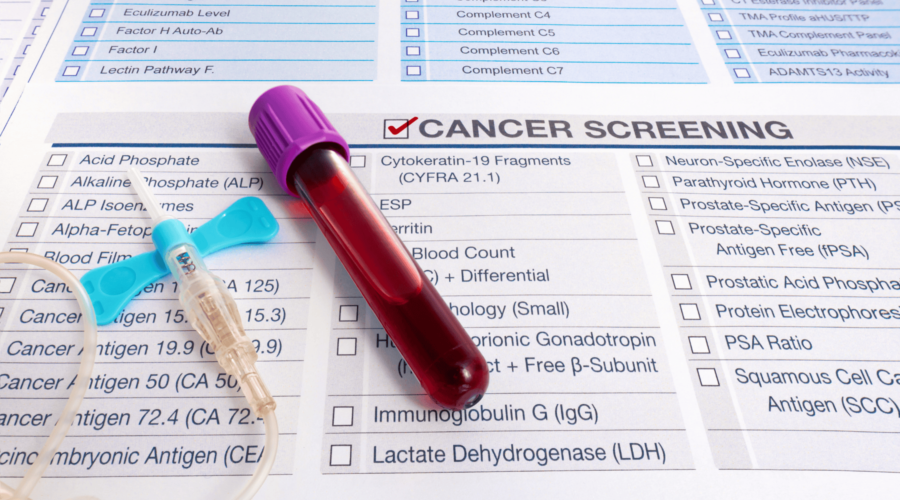
Blood-based markers such as CA-125 and HE4 are the most widely used, but others such as LDH (lactate dehydrogenase), β-hCG, AFP (alpha-fetoprotein), and CEA may provide clues depending on the subtype.
These biomarkers, rooted in ovarian cancer pathophysiology, help doctors track disease activity, detect recurrence, and personalise ovarian cancer treatment strategies.
Imaging Correlations
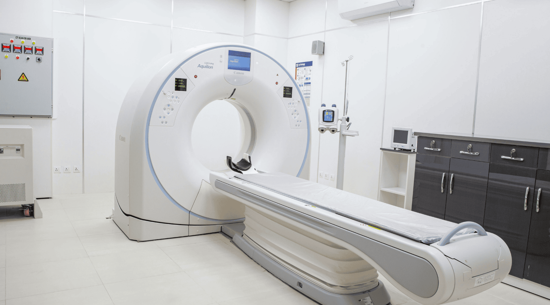
Imaging tools, like ultrasound, CT, and MRI, don’t just spot masses; they reveal patterns consistent with epithelial ovarian cancer pathophysiology. Features such as irregular solid areas, ascites, or peritoneal nodules align with known pathways of spread, guiding both surgical planning and therapeutic decisions.
Cytoreduction Relevance
Cytoreduction (or debulking) means removing as much tumour tissue as possible. The reason is clear from ovarian tumour pathophysiology: residual cancer cells fuel recurrence and worsen survival outcomes. Evidence consistently shows that patients with little to no visible disease after surgery have the best prognosis.
Targeted Therapies (PARP, VEGF inhibitors)

PARP inhibitors exploit DNA repair defects, especially in BRCA-mutated tumours, turning genetic weakness into a therapeutic advantage. VEGF inhibitors, on the other hand, target abnormal blood vessel growth that drives ascites and tumour progression. They’re a great example of applying pathophysiology of ovarian cancer to the treatment.
Why understanding biology can change everything
The pathophysiology of ovarian cancer is intricate, weaving together genetic mutations, immune evasion, angiogenesis, and ascites formation. What’s surprising is that researchers are also uncovering links between ovarian cancer and hormonal influences, like estrogen signalling, which may help explain why the disease behaves differently across life stages.
Anyone touched by ovarian cancer should know: there’s a community, a care team of oncology doctors, and knowledge standing beside you.
Understanding ovarian cancer pathophysiology isn’t just medical theory: it’s a way to demystify the disease, ask the right questions, and feel more in control.
FAQs on pathophysiology of ovarian cancer
It refers to the biological processes, like genetic mutations, cell growth changes, and spread patterns, that drive how epithelial ovarian cancer develops and progresses.
Related Blogs


Nutrition5 min read
(Protein powder ke fayde): प्रोटीन पाउडर के फायदे और इसे लेने का सही तरीका
Dr. Vrundali Kannoth