

Education5 min read
Vitamin B1 Thiamine Supplements: Nerve Function & Cancer Recovery Support
Dr. Vrundali Kannoth
If you or someone you love has recently had surgery, noticing an unexpected swelling or fluid buildup near the incision site can feel unsettling. You might wonder whether something has gone wrong or if your stitches are coming apart.
While it can cause worry, there's no need to feel that way. A seroma is a common part of post-surgery healing where fluid builds up under your skin. It often heals on its own, and in some cases, you may need additional support from your care team.
However, understanding what a seroma is and how it develops can help ensure you reach out to your care team at the right time.
In this guide, we'll cover seroma meaning, what causes it, how to identify one, and the treatment options available to you.
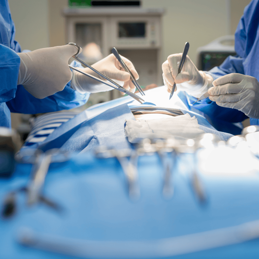
A seroma refers to a buildup of serous fluid that collects in a pocket under the skin. This pale-yellow liquid is something your body naturally produces, and it tends to gather in areas where tissue has been removed. It can also be considered one of the very common side effects of cancer surgery.

Think of it as your body's way of responding to the space left behind after a procedure. The fluid isn't blood or pus.
A seroma can range from a tiny, barely noticeable pocket to a larger, more visible swelling. It may feel like a soft, fluid-filled lump near your surgical site. The good news is that most resolve on their own with time.
Some may need medical attention depending on their size and location, but either way, your care team will know exactly how to help.
Seroma formation typically occurs after any procedure that involves cutting, lifting, or removing tissue. When the body detects that open space, it sends fluid to the area as part of its natural effort to heal.
Here are some of the most common causes:
None of these causes is unusual or a sign that something went wrong. They're simply part of how the body responds to surgery. Being aware of your personal cancer risk factors and sharing your full medical history with your surgeon can also help anticipate these kind of potential complications.
Not all seromas look or behave the same way. Here's a closer look at the most common types.
A small seroma is often the least concerning type, and it often resolves on its own within a few weeks. It usually shows up as a minor, soft swelling near the incision and may be barely noticeable. Many people find it only when they touch the area during routine wound care.
A chronic seroma persists for weeks or even months after surgery and doesn't heal on its own. It may continue to refill even after being drained.
Sometimes it can develop a thickened wall of tissue around it, making natural reabsorption more difficult. Your doctor may recommend more active seroma management techniques if this develops.
A calcified seroma feels firm or solid rather than soft and fluid-filled. It may sometimes be mistaken for a seroma lump or even a tumour on imaging, which is why accurate cancer diagnostics are so important.
Though usually harmless, it may require removal if it causes discomfort or diagnostic confusion.
A residual seroma refers to a small amount of fluid that remains after the main pocket has mostly resolved or been treated. It's the leftover fluid your body hasn't yet fully cleared.
Most residual collections resolve with time and patience, though monitoring ensures nothing else develops in their place.
A seroma cyst is a fluid pocket that becomes enclosed by a fibrous capsule, creating a defined, contained structure. This happens when your body walls off the fluid as part of its natural recovery response.
If left alone, a seroma cyst can persist and may need a minor procedure for removal. Your surgical oncology team can help decide the best course of action for seroma healing.

In most cases, a seroma is not dangerous, but still letting your care team know will not hurt you. Since you're recovering from your surgery, they can ensure that your seroma doesn't worsen or cause problems in your recovery. Here's how you can identify one:
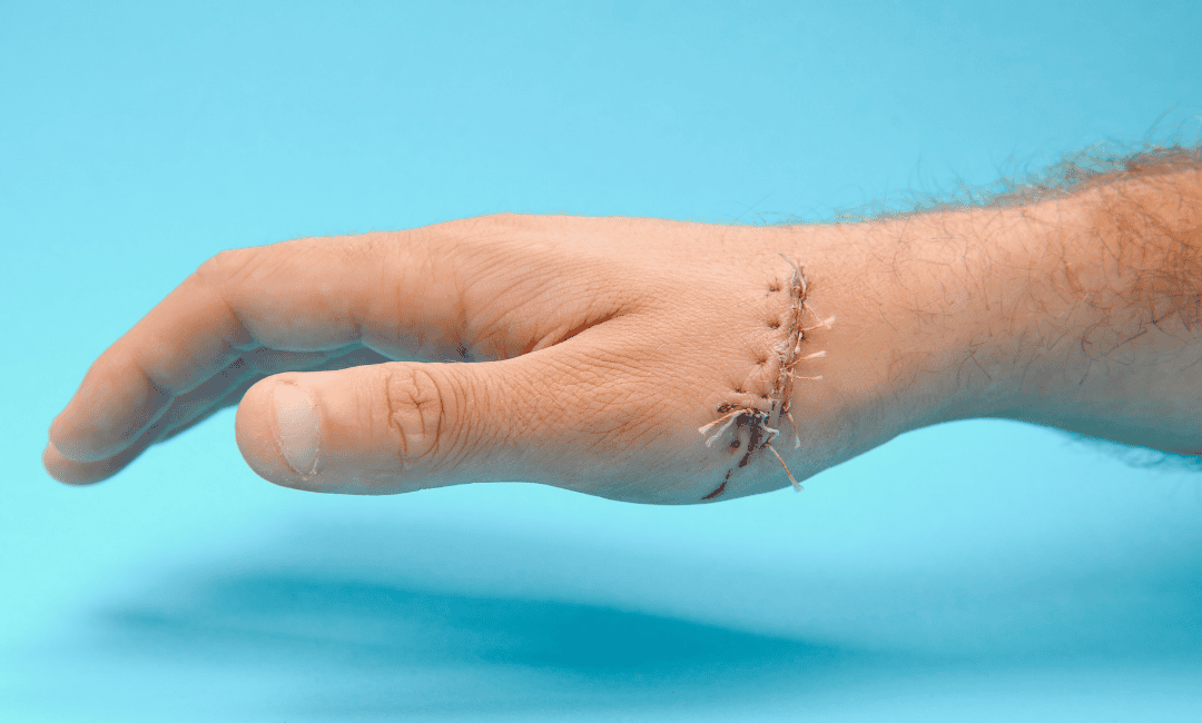
One of the earliest signs is a visible swelling or bulge near your surgical site, appearing days or even weeks after the procedure. A soft tissue seroma like this usually isn't accompanied by redness or warmth, which helps distinguish it from an infection.
When you gently press the swollen area, it may feel like a soft, smooth water balloon beneath the skin. This sensation is normal and not a cause for alarm, but do consider its size and share the details with your oncology doctors.
Sometimes, seroma fluid may leak through the incision line as a clear or light-yellow discharge. This is different from the cloudy or foul-smelling drainage linked to an infected seroma, but consistent or heavy leaking is a sign to reach out to your doctor.
Keeping the area clean and dry remains important during this time.
Some people experience seroma pain, which can feel like tightness, pressure, or mild aching that increases as the fluid pocket grows. While it isn't usually severe, persistent pain should always be discussed with your doctor as part of your overall cancer treatment plan.
While a seroma post surgery can happen to anyone, certain factors may increase the likelihood. Knowing these risk factors helps you and your healthcare team stay one step ahead and plan for a smoother recovery.
Here are some of the most common risk factors to be aware of:
Every person heals at their own pace, and having one or more of these factors doesn't mean complications.
Usually, the seroma heals on its own because your body will reabsorb the fluid. This takes at least a few weeks to months. Your doctor may suggest wearing a compression garment to apply gentle pressure and encourage the absorption process.
In other cases where it does not heal naturally, you might need the following seroma treatment depending on its size, symptoms, and presence:
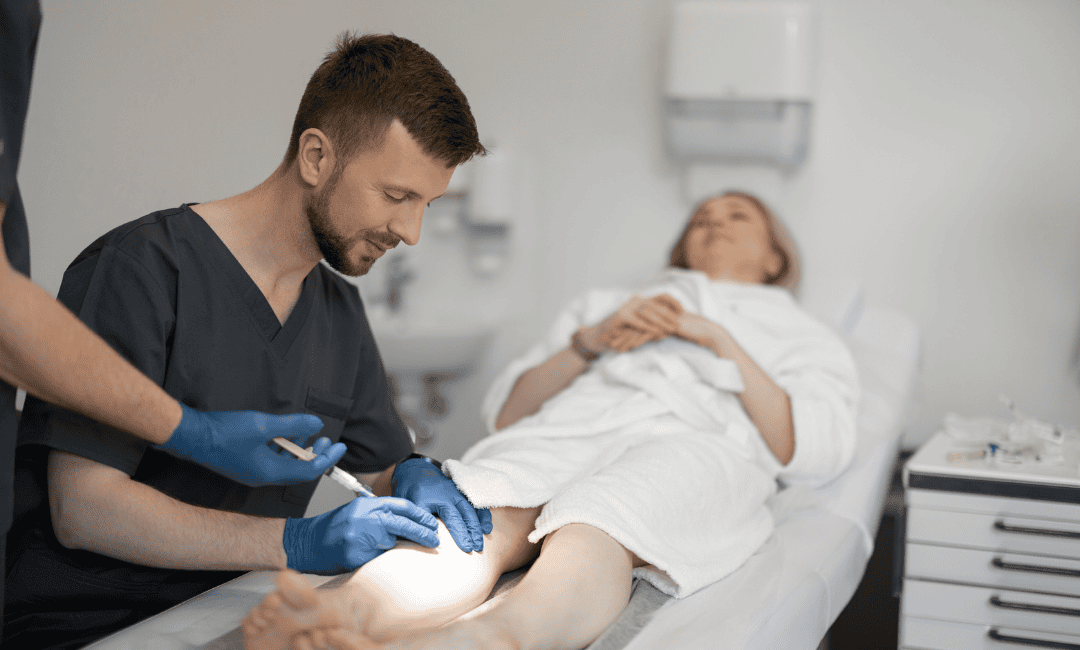
If your seroma is large and leaking, your doctor may perform seroma aspiration, where a needle is used to drain the fluid gently. It's a quick, simple process, though some seromas may refill and need repeat drainage.
For seromas that keep returning, a solution is injected into the cavity to encourage the walls to seal together. This collapses the space so fluid can no longer collect there. It’s an effective seroma treatment for stubborn seroma collection.
In rare cases, your care team may discuss how to remove seroma surgically by taking out the fibrous capsule and closing the cavity. This is only considered when other approaches haven't worked, and seroma complications like this are uncommon.
Dealing with a postoperative seroma can be frustrating, especially when you've just had surgery. But know that this is a common, natural, and most importantly, highly manageable condition.
The most essential thing you can do is stay informed and connected with your care team. Don't hesitate to reach out if something even slightly feels off. In most cases, you won't even need a needle or any additional procedure.
Just trust your body's ability to heal, follow your postoperative instructions carefully, and rest well after your surgery. Recovery is not always a straight line, but with the right support and knowledge, you can ensure a more smooth journey.
At EverHope, our experts are here to make your healing as comfortable as possible. We offer not just advanced medical treatments but well-rounded support that puts your well-being at the centre of everything we do.