
+917795060087
Call Us
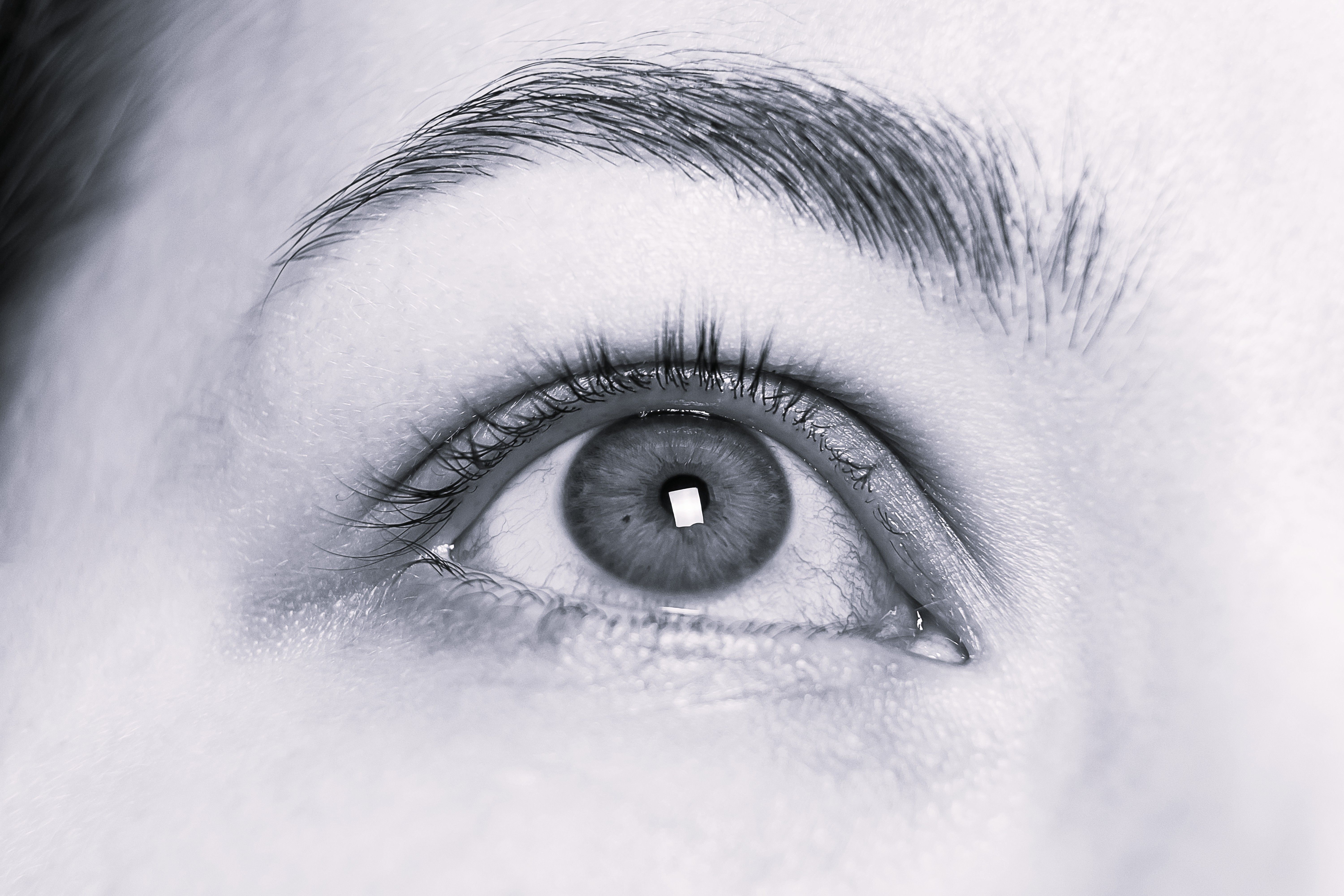
Eye Cancer
What is Eye Cancer?
Eye cancer refers to the malignant tumors that arise or occur near the eye. The eyelids and tear glands ( adnexa), orbit ( the tissue that surrounds the eyeball), or eyeball( globe) can be involved. Survival and vision preservation are possible with early detection, and hence, early detection is highly encouraged.
Eye Cancer Types
Retinoblastoma
•A rare but aggressive form of cancer that originates in the retina, most frequently occurring in children younger than 5 years old. Symptoms can include a white reflex within the pupil or crossed eyes. Treatment is chemotherapy, laser treatment, cryotherapy, or even the removal of the eye in very advanced stages. Early diagnosis can salvage both vision and life.
Uveal Melanoma
•The most frequent primary intraocular cancer in adults that develops from the pigmented uveal tract (iris, ciliary body, or choroid). It usually develops symptomlessly but may lead to loss of vision, floaters, or flashes of vision. Treatment options may include radiation therapy (brachytherapy or proton beam therapy), laser treatment, or removal of the eye in extreme cases.
Ocular Surface Squamous Neoplasia (OSSN)
•A tumor of the conjunctiva or cornea, which commonly occurs in sun-exposed persons or immunocompromised patients. Presents as a white or pink swelling on the surface of the eye. Topical chemotherapy, surgical removal, or cryotherapy is used to treat it. Follow-up is crucial because there is a tendency to recur.
Sebaceous Gland Carcinoma
•A malignant and very rare tumor derived from oil glands in the eyelid. It can masquerade as a benign eyelid infection, leading to delayed diagnosis. It presents with a painless swelling of the eyelid, loss of eyelashes, or swelling of the eyelid. Surgery is the typical treatment and radiation may be added if the tumor is large.
Lymphoma of the Eye
•Usually involves the conjunctiva, orbit, or uvea and is related to systemic lymphoma. Symptoms include painless swelling, proptosis, or diplopia. It is diagnosed through biopsy. Radiation and chemotherapy are usually used based on the type and extent of disease.
Eye Cancer Symptoms
What’s Notable
The most common eye cancer in children is retinoblastoma, and the most common one in adults is melanoma.
Certain eye cancers are even detectable through a routine eye checkup before any symptoms are felt.
Exposure to UV radiation for many years, particularly without protective eye cover, increases the risk of ocular melanoma and surface eye cancers.
When to Seek Help
Key signs of an eye tumor include sudden vision changes, visible dark spots or bulging, persistent pain or redness, flashes or floaters, loss of side vision, and unexplained tearing or swelling. These symptoms should be promptly checked by an eye specialist.
Eye Cancer Causes & Risk Factors
Age and Heredity
Retinoblastoma occurs in early childhood, but melanoma and lymphoma of the eye occur in most older people. Gene mutations that are hereditary, such as RB1, are unsafe.
Sunlight and UV Exposure
Exposure to UV radiation for many years, particularly without protective eye cover, increases the risk of ocular melanoma and surface eye cancers.
Fair Skin and Light Eyecolor
Individuals with blue, green, or gray-colored eyes and light skin coloured are at increased risk for developing intraocular melanoma.
Family History of Eye Cancer
A first-degree relative with retinoblastoma, uveal melanoma, or other eye cancer suggests a genetic component and should prompt early evaluation.
Immune Suppression and HIV/AIDS
Those with compromised immunity are at increased risk for developing eye lymphomas or eye Kaposi sarcoma.
Exposure to Specific Chemicals
Occupational exposure to welding fumes, pesticides, or solvents may be associated with greater risks of eye cancer.
Eye Cancer Diagnosis
Symptom Awareness & Initial Check-up
Step 1: Symptom Awareness & Initial Check-up
Blurred vision, partial blindness, eye pain, or an increasing dark spot should lead to an immediate eye test by an ophthalmologist.
Eye Cancer Treatment & Therapy
Chemotherapy
What it does:
Kills rapidly growing bladder cancer cells throughout the body.
Treated for:
Retinoblastoma (especially if advanced); metastatic disease.
Recovery:
Taken in cycles; can be tired, nauseated, and have risk of infection.
Targeted Therapy & Immunotherapy
What it does :
Specifically targets mutations or boosts immune function to fight cancer.
Treated for:
Advanced uveal melanoma or metastatic eye cancer.
Recovery:
Side effects are unforeseen; usually less than with chemotherapy; must be closely monitored.
Management & Prevention
Vision Monitoring
•Periodic eye checkup and screening for vision change and tumor control.
Post-Treatment Care
•Protection of eyes and lubricating drops after surgery or radiation, and healing ointments.
Rehabilitation Support
•Visual aids such as magnifying glasses or prosthetic eyes can help restore independence and enhance quality of life following loss of an eye or blindness.
Why Choose Everhope For Eye Cancer?
At Everhope, our dedicated team supports your journey with advanced care, compassionate guidance, and lasting hope.
2.5K
New ocular melanoma cases reported annually in US.
2.3K
New retinoblastoma cases reported in India in 2022
1 out of every 18,000
live births in India develops retinoblastoma at birth.
Explore Our Latest Updates
Insights5 min read
Muh Ke Cancer Ke Lakshan (मुंह के कैंसर लक्षण): कैसे पहचानें?
Dr. Vrundali Kannoth

FAQs on Eye Cancer
No question is too small when it comes to your care
Yes, the majority of eye cancers can be cured—especially if discovered at an early stage. The chance of cure depends on the type, size, and site of the tumor.
Find a Centre Near You
Gurgaon EBD 65
EBD 65, Sector 65, Golf Course Extension Road, Gurgaon