

Symptoms5 min read
Folic Acid Supplement: Importance, Benefits and Deficiency Symptoms
Dr. Vrundali Kannoth

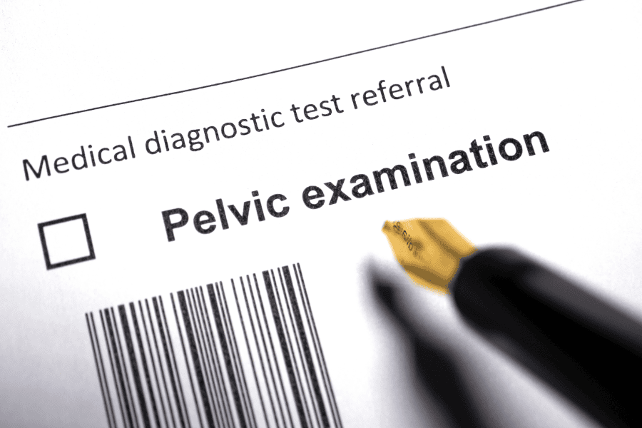
Your doctor felt something unusual during your pelvic examination, or perhaps an ultrasound for unrelated symptoms revealed an unexpected finding described as a mass in pelvic area.
Now you're sitting with test results in hand, feeling anxious about what this discovery means and whether it's something serious requiring urgent treatment. The term "pelvic mass" sounds ominous, bringing immediate worry about cancer even though your doctor mentioned most masses turn out to be benign.
Understanding what is a pelvic mass and learning about the various causes helps put your situation into proper perspective.
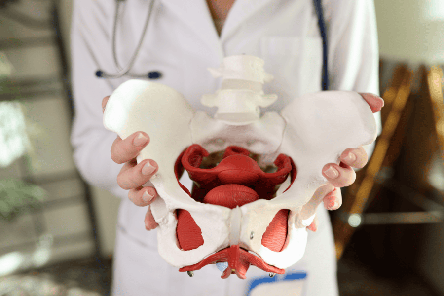
Pelvic mass meaning describes any abnormal growth or collection of tissue located in the pelvic cavity between your hip bones, which your doctor can feel during examination or detect through imaging studies.
A pelvic mass female presentation differs substantially from pelvic mass male because women's reproductive organs represent the most common mass sources, while men's pelvic masses more often originate from the prostate, bladder, or gastrointestinal tract.
According to research, approximately 70-80% of pelvic masses detected in premenopausal women represent benign conditions like ovarian cysts or uterine fibroids.
Right pelvic mass versus left-sided masses show different patterns in frequency and common causes, though overlap exists considerably.
| Feature | Right pelvic mass | Left pelvic mass |
|---|---|---|
| Common ovarian causes | Functional cysts from normal ovulation cycles | Functional cysts from normal ovulation cycles |
| Gastrointestinal structures | Appendix (appendiceal mucoceles, appendiceal cancer), cecum, terminal ileum | Sigmoid colon (diverticular masses, colonic lesions) |
| Typical presentations | Appendix-related masses, right ovarian pathology, cecal tumours | Sigmoid diverticulitis, left ovarian pathology, descending colon masses |
| Kidney involvement | Right kidney pathology (less common as pelvic mass) | Left hydronephrosis, left kidney tumours extending into pelvis |
| Clinical consideration | Appendicitis must be ruled out with acute right-sided pain | Diverticulitis common differential with left-sided symptoms |
Pelvic mass location significance: While location provides diagnostic clues, it doesn't definitively determine whether a mass is benign or malignant.
Central pelvic masses typically arise from the uterus or bladder, whilst lateral masses more commonly originate from ovaries, fallopian tubes, or bowel structures.
The causes of pelvic mass span a wide spectrum from completely harmless functional cysts to serious malignancies requiring urgent treatment.
Research emphasises that systematic evaluation effectively narrows diagnostic possibilities based on patient age, symptoms, examination findings, and imaging characteristics.
The symptoms range from completely asymptomatic (discovered incidentally) to causing severe symptoms requiring emergency treatment. Here are some of the common sympotoms:
Pelvic or abdominal discomfort: Dull, aching pain or pressure in the lower abdomen represents the most frequent symptom. Sharp, sudden pain suggests complications like ovarian cyst rupture or torsion (twisting).
Urinary symptoms: Frequent urination occurs when masses press on the bladder. Difficulty emptying bladder completely or urinary urgency develop with larger masses. Blood in urine (hematuria) suggests bladder involvement or severe compression.
Bowel symptoms: Constipation, bloating, or difficulty with bowel movements result from masses compressing the rectum or colon. Abdominal lump visible or palpable by the patient sometimes occurs with very large masses.
Menstrual changes: Irregular bleeding, heavy periods, or bleeding between periods can accompany uterine or ovarian masses. Postmenopausal bleeding always requires evaluation as it suggests potential malignancy.
Cancer symptoms that warrant urgent evaluation: Unexplained weight loss exceeding 5% of body weight without dietary changes raises concern for malignancy.
Moreover, persistent bloating or early satiety (feeling full quickly) are classic ovarian cancer symptoms often dismissed initially. Severe, acute pain may indicate complications requiring emergency surgery.
So, how often are pelvic masses cancerous? That’s a question that concerns most patients. Reassuringly, only 5-10% of masses in premenopausal women are malignant, though risk increases to 20-30% in postmenopausal women. However, pelvic mass male presentations carry higher malignancy rates.
Understanding different pelvic mass types helps doctors predict what they're likely dealing with and plan appropriate next steps.
Fluid-filled (cystic) masses: Most benign pelvic masses are cysts containing fluid.
Solid pelvic mass types: When a mass is completely solid tissue rather than fluid-filled, doctors pay closer attention because cancer risk is higher. That said, fibroids (extremely common benign tumours) are solid but almost never cancerous. Solid ovarian masses need careful evaluation to rule out cancer, as do enlarged lymph nodes or other solid growths.
Mixed masses: Some masses contain both solid and cystic parts, which makes diagnosis more challenging. Dermoid cysts, for example, look complex on scans but are typically benign, while some ovarian cancers also appear as mixed masses, so further testing becomes essential.
From reproductive organs: About 70-80% of pelvic masses in younger women originate from ovaries, fallopian tubes, uterus, or cervix. These are gynaecological masses.
From other pelvic structures: Some masses arise from your bowel (colon or appendix), urinary system (bladder or kidney), or other structures like lymph nodes or blood vessels. These become more common after menopause and account for most pelvic mass lesion cases in men.
Understanding these categories helps your doctor determine whether you need immediate surgery, can wait and monitor with follow-up scans, or might benefit from medical treatment first.
Cancer diagnostics for pelvic masses involve systematic evaluation combining clinical assessment, imaging studies, and sometimes tissue sampling.
The treatment depends on mass characteristics, patient age, symptoms, and cancer likelihood rather than following one standard approach.
Observation:
Medical management:
Surgery for benign masses:
Cancer treatment:
Research shows that benign masses often need only simple interventions, but malignancies require multidisciplinary teams coordinating comprehensive cancer treatment.
Discovering a pelvic mass understandably triggers anxiety, but remember that most pelvic masses in premenopausal women represent benign conditions like functional cysts or fibroids requiring minimal intervention.
However, even when masses need treatment, many options exist, ranging from simple observation to minimally invasive surgery.
The key lies in not dismissing pelvic mass symptoms that persist, working with your healthcare team to establish a proper diagnosis, and following through with recommended monitoring. Early evaluation of concerning masses leads to better outcomes, whether the cause is benign or malignant.
For a comprehensive evaluation of pelvic masses, connect with experienced gynaecologists and oncology specialists who can provide personalised assessment and care tailored to your individual needs.